AI tool uses face photos to predict cancer survival
Researchers in the US have developed an AI foundation model – trained on millions of facial images – that seems to be able to directly predict survival risk in patients with cancer.
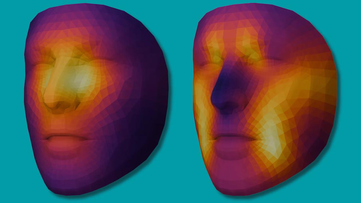
The team from Mass General Brigham has published a paper in The Lancet Digital Health that describes two algorithms in the model, one that estimates biological age (FAHR-FaceAge) and a second (FAHR-FaceSurvival) that generates the survival prediction forecast.
Their study found that FAHR-FaceAge could identify biological age with an error margin of around five years – which the researchers say offers greater accuracy than other tools – and that the deviation between a patient's FAHR-FaceAge and their chronological age was independently linked to survival.
FAHR-FaceSurvival was designed to go a step further by learning and predicting survival from a single face photograph directly, rather than estimating it based on biological age.
"To our knowledge, this is the first demonstration that an AI model can directly predict cancer survival from a simple photograph," said Hugo Aerts, co-senior author and director of the AI in Medicine (AIM) programme at Mass General Brigham.
The algorithms rely on different parts of the face to make predictions of age and mortality risk, according to the paper. FAHR-FaceAge focuses on the nasolabial folds, the forehead area just above the orbits and the temples, while FAHR-FaceSurvival evaluates the region below the eyes and around the nasal bridge.
The algorithms were trained on more than 40 million unlabelled facial images across a range of ethnicities and ages from the WebFace42M dataset, developed by researchers from Tsinghua University, Imperial College London, and XForwardAI, and were validated on a panel of nearly 5,000 patients with cancer treated with radiotherapy in the Netherlands.
Unlike other models relying on specialised imaging, the FAHR-Face algorithms used consumer-grade facial photographs, which should broaden their clinical applicability, the study authors write in the paper.
"As the population ages, physicians need better ways to estimate physical reserve and frailty before recommending intensive treatments such as radiation, surgery, or systemic therapy," said corresponding author Raymond Mak, a radiation oncologist at Mass General Brigham Cancer Institute and AIM faculty member.
"Our goal is to make face photograph-based health assessment more quantitative, consistent, and scalable so it can support personalised treatment decisions rather than replace physician judgment."
The team is also expanding this research through the FaceAge Research Study, which is open to the public at faceage.bwh.harvard.edu.
Participants can upload a facial photograph and receive their own FaceAge research readout, while also contributing data that will help researchers improve and validate AI-based measures of facial ageing and health across larger and more diverse populations.
Prof Miroslaw Bober, an imaging and AI specialist at the University of Surrey in the UK, said that the study is "interesting and substantial" and showed that the tools can separate patients by risk.
That said, he added that the study "did not test whether the AI recognises anything a doctor would not. It has not shown that these predictions add clinically useful information beyond doctors' assessments of a patient's general health and fitness, which were not measured in the study. This is a promising proof of concept, but it is not ready for clinical use."
Photo by Cristina Zaragoza on Unsplash