Solution Spotlight: Modelling human myelin and predicting repair
Demyelinating diseases, including multiple sclerosis (MS) and Charcot-Marie-Tooth (CMT) disease, present a substantial and escalating burden. Each of these conditions afflicts approximately three million individuals worldwide.
Notably, the prevalence of MS has seen a sixfold increase over the past four decades, a trend that has not been matched by corresponding advances in therapeutic efficacy. In addition, the failure rate for disease-modifying interventions exceeds 99%, underscoring a persistent gap between clinical need and available treatment modalities. Epidemiological data further delineate the scope of the problem, with chronic inflammatory demyelinating polyneuropathy and Guillain-Barré syndrome contributing additional patient populations to this landscape.
Technical and biological impediments in myelinating cell research
The study of myelinating cell types is encumbered by several fundamental challenges. Chief among these is the difficulty in establishing and maintaining functional populations of myelinating cells in vitro.
Three principal obstacles are routinely encountered. First, animal models exhibit species-specific divergences in myelin biology, thereby limiting the translational applicability of preclinical findings. Second, human oligodendrocytes and Schwann cells have historically proven recalcitrant to derivation and maintenance in culture. Third, conventional two-dimensional culture systems lack the spatial architecture requisite for authentic myelin wrapping, further constraining the physiological relevance of experimental models.
3D organoids: Addressing species and technical barriers
In response to these challenges, 28bio has developed organoid technologies that incorporate mature, functional myelinating cell types within a three-dimensional architecture.
Both CNS-3D and PNS-3D organoids support authentic myelin formation and enable high-throughput measurement of myelination for therapeutic screening. The CNS-3D platform generates oligodendrocyte-myelinated brain organoids containing neurons, astrocytes, and oligodendrocytes, while the PNS-3D platform establishes Schwann cell-myelinated nerve organoids comprising Schwann cells and neurons. This configuration recapitulates both central and peripheral nervous system myelination, thereby overcoming both technical and species-specific barriers.
Oligodendrocyte differentiation and developmental fidelity in CNS-3D
The differentiation protocol for CNS-3D myelination organoids contains oligodendrocyte precursors and mature myelinating oligodendrocytes, in addition to neurons and astrocytes. Temporal marker progression within these organoids mirrors published human oligodendrocyte developmental timelines, supporting their physiological relevance.
The developmental sequence proceeds from neural stem cells to neurons and astrocytes, followed by the emergence and maturation of oligodendrocyte precursors. Marker expression analysis over an 84-day period demonstrates that early markers plateau by week six, while oligodendrocyte markers exhibit delayed onset and progressive augmentation through week twelve.
Molecular and structural verification of authentic myelination
Functional myelin formation within these organoids is substantiated by multiple analytical modalities.
Quantitative PCR analysis reveals that myelin basic protein (MBP) expression increases substantially from week eight to week 12, indicating active myelin synthesis concurrent with oligodendrocyte maturation. Immunofluorescence imaging confirms the spatial colocalisation of MBP with neurofilament-positive axons at week 12, demonstrating structural myelin formation. This spatial arrangement signifies authentic myelin wrapping around axonal structures, rather than non-specific protein accumulation, thereby validating the physiological relevance of the organoid system.
Pharmacological validation using Clemastine
The technologies' utility is further demonstrated through pharmacological validation with Clemastine, a promyelinating compound possessing established clinical trial activity.
Clemastine enhances MBP expression in a manner proportional to treatment duration. Quantitative PCR analysis demonstrates treatment-dependent upregulation, with maximal response observed at day 84 following a four-week exposure. Western blot analysis corroborates these findings at the protein level. The dose- and duration-dependent response to Clemastine showcases the platform’s capacity for identifying compounds that modulate myelin disruption or repair, establishing its suitability for therapeutic screening.
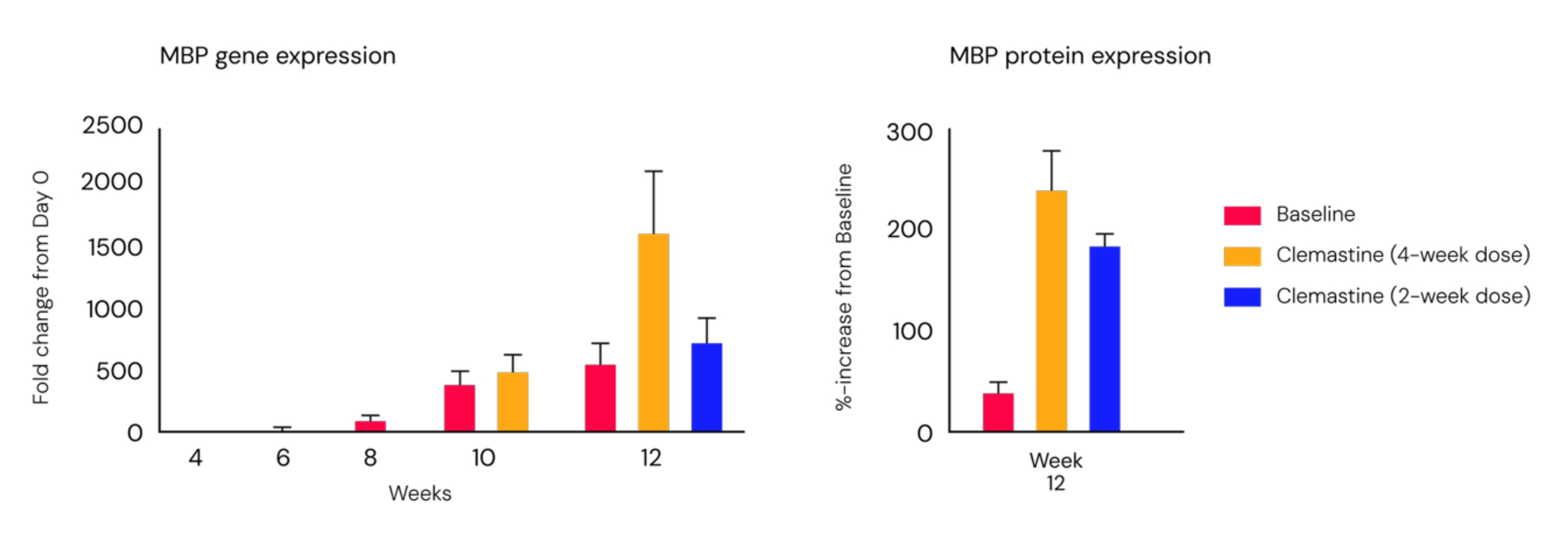
(Left) qPCR geneanalysis demonstrates treatment-dependent MBP upregulation, with maximum response at Day 84. (Right) Western blot confirms corresponding protein-level changes, demonstrating pharmacological modulation at both molecular and protein levels.
Schwann cell integration and functional nerve establishment in PNS-3D
In the PNS-3D system, the establishment of myelinated, functional nerve organoids is contingent upon the integration of primary human Schwann cells. These cells align with neurons and generate myelin sheaths, as confirmed by fluorescent microscopy.
Transmission electron microscopy highlights the presence of compact myelin ultrastructure around individual axons. Functional analysis reveals that cultures containing Schwann cells exhibit approximately 100 functional responses, whereas cultures lacking Schwann cells demonstrate fewer than 10 responses. This disparity confirms the necessity of myelinating cells for baseline nerve function for therapeutic assessment.
Comprehensive analytical endpoints for phenotypic and mechanistic investigation
Both CNS-3D and PNS-3D myelinating organoids enable phenotypic screening and mechanistic investigation through a suite of quantifiable endpoints. Functional analysis, including electrophysiological assessment of network activity, is available in both technologies.
Quantitative PCR and Western blot analyses provide molecular and protein-level validation of myelination pathways. Immunohistochemistry offers qualitative fluorescent labelling for spatial analysis, with CNS-3D fully established and PNS-3D at baseline. Degeneration structural analysis via quantitative phase imaging is available in PNS-3D. These endpoints collectively facilitate a comprehensive evaluation of both mechanistic and phenotypic effects.
Mechanistic and phenotypic screening for demyelination and repair
CNS-3D and PNS-3D myelinating organoids provide physiologically relevant models for the investigation of myelin disruption and repair. These technologies enable both mechanistic investigation through molecular and structural endpoints and phenotypic screening through functional assessment. Their dual capability positions them for the identification of therapeutic candidates that promote remyelination or prevent demyelination.
By addressing the species-specific limitations of animal models and the architectural constraints of two-dimensional cultures, these human-derived, three-dimensional systems offer a gateway to the advancement of therapeutic development in demyelinating diseases.
To learn more, please view "Modeling Myelin Disruption and Repair for Therapeutic Assessment."
About 28bio
28bio is a neurotechnology company engineering human brains at-scale exhibiting memory, learning, and cognitive functions. Its Nexon™ platform integrates tissue engineering, neural interfacing, and AI to reverse today’s neurological health crisis by improving the ability to predict which therapies will work in humans. 28bio is committed to advancing ethical standards in the development of brain organoid technology and engineered human cognition. For more information, visit 28bio.com.