Recent breakthroughs in 3D bioprinting: A look at trends and challenges

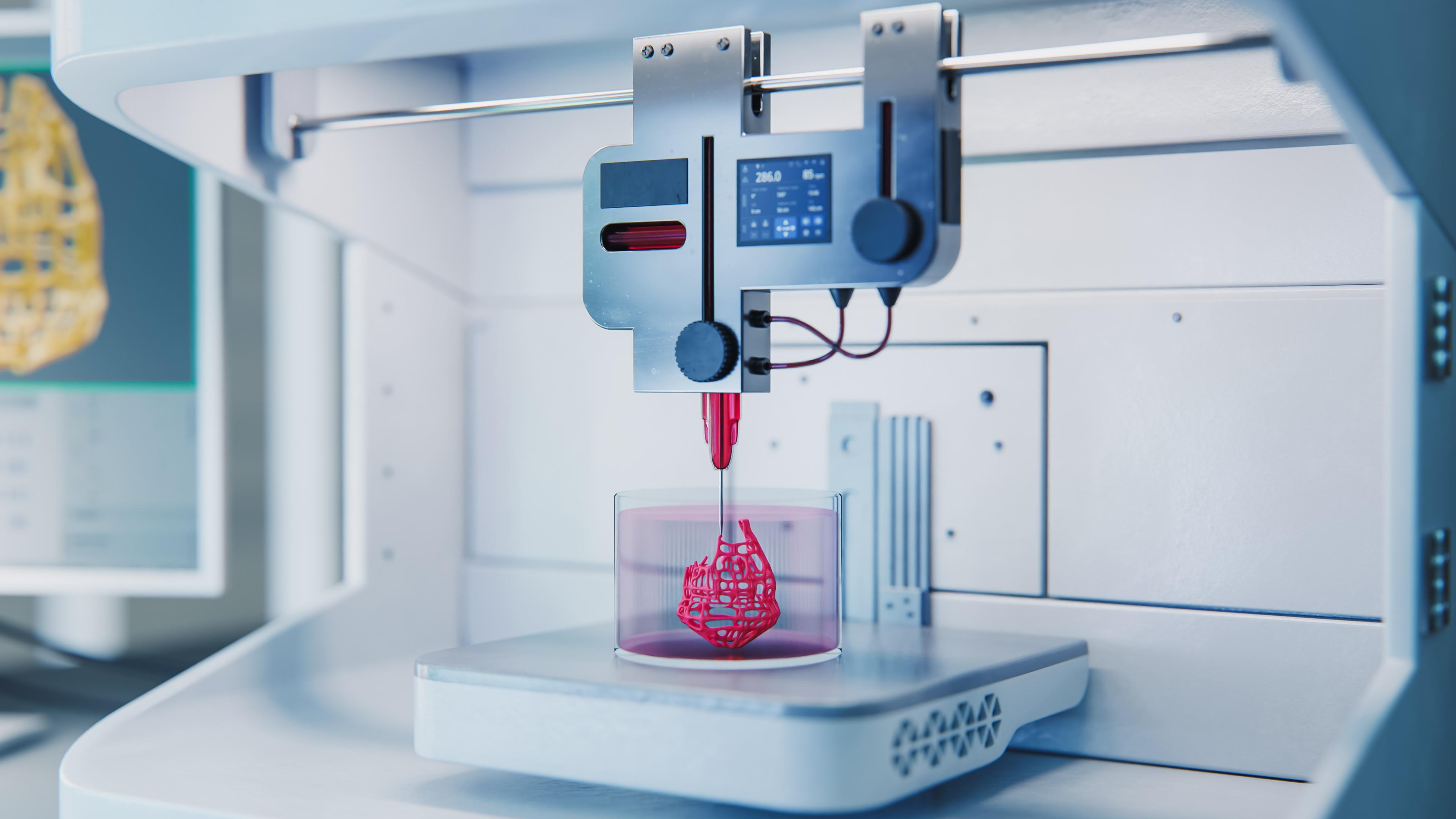
3D bioprinting technology is bringing a new revolutionary wave to the healthcare industry.
This technology combines biomaterials, bioinks, and 3D printing to develop specific structures that mimic functional tissues or organs. Currently, these bio-printed models are used as a drug screening and discovery tool to accelerate the production of potential drugs. However, researchers are continually focusing on the development of bioinks with improved mechanical properties, better printability, and biocompatibility to mimic the natural extracellular matrix of tissues.
The recent breakthrough includes the introduction of elastic hydrogel materials, cellular matrices, and incorporation of functional cells and tissues, such as autologous mesenchymal stem cells for the reconstruction of bones. This elastic hydrogel material is specifically designed for 3D printing of soft living tissue, like blood vessels. Additionally, cellulose-based inks are also gaining traction owing to their versatility in drug discovery, tissue engineering, bone reconstruction, and regenerative medicine.
As researchers progressively explore 3D bioprinting across various healthcare applications, the transformatory innovation in this field is clear.
3D bioprinting: Current landscape
3D bioprinting offers precise control to print tissue structures that can act fully functionally for regenerative medicine, as bond bandages to heal wounds, and in weight-bearing bone reconstruction and skin grafts to address the specific challenges encountered during foot and ankle surgery, due to accidental injuries and tumour resection or Charcot foot, for example. Although traditional bone grafting approaches, such as allografts, autografts, and bone substitutes are available, there are certain limitations, such as morbidity, donor site pain, and variability rates of resorption that may hinder the treatment.
3D bioprinting fundamentally involves an approach of bioinks deposition in a spatial manner to create a 3D structure that works like bone tissue. Several bioprinting techniques, such as extrusion-based bioprinting, inkjet-based bioprinting, and stereolithography-based bioprinting poses the ability to fabricate scaffold with controlled pore size and interconnectivity, which is essential for nutrient diffusion and vascularisation. Therefore, the precise incorporation of multiple cell types and growth factors with scaffolding, mimicking the biochemical environment of bone tissue, holds an immense opportunity for personalised bone reconstruction in the ankle and foot.
Current research on weight-bearing bone reconstruction
Several preclinical studies are presently demonstrating the feasibility of bioprinting bone constructs with improved mechanical strength and osteogenic potential for load bearing applications. For instance, in a recent study scientists successfully showed the development of biomimetic structural design in 3D-printed scaffolds for bone tissue engineering and successfully optimised key parameters in biosensitive materials, such as tissue connectivity, porosity, pore size, and mechanical strength.
This structure mimics the functional complexities and structural integrity of native bone, thereby enhancing the mechanical performance for weight-bearing capabilities. Furthermore, the study highlighted the integration of advanced technologies, such as CAD and AI, enabling the design of patient-specific scaffold design for weight-bearing bone construction. In case of significant bone loss due to trauma and injuries, these bioprinted constructs can help to fill large bone defects to restore biomechanical function.
Recent case study of 3D bioprinting and bone defect correction
Bioprinting technology has given a new direction to the healthcare industry, allowing treatment of patients with significant bone defects due to accidental injuries.
A case study in the Journal of Orthopedic Case Reports highlighted the successful treatment of a 38-year-old male patient with critical-size bone defect with 3D printed mesh implants. Initial treatment was done by doctors with fibula plating and an external fixator. However, the fracture still showed atrophic non-union and 2.5 cm limb shortening, so a team of doctors operated with 3D printed titanium mesh implants with plate constructs. After 1.5 years, a good bone integration and ambulation restoration could be seen in the patient’s CT scan.
Top trends in 3D bioprinting technology
The field of 3D bioprinting technology is witnessing rapid advancements, offering new possibilities in regenerative medicine, tissue engineering, and pharmaceutical research. Some of the trends that are must to watch in 3D bioprinting market are listed below.
1. Stimuli-responsive biomaterials
Industrial players are making significant progress in creating novel biomaterials and bioinks. The focus will be on the development of stimuli-responsive biomaterials with improved biocompatibility, mechanical strength, printability, and the ability to mimic tissue environment.
For instance, collagen, alginate, gelatin, and decellularised extracellular matrix are currently being refined to improve structural integrity and cellular interaction. Furthermore, cellulose-based bioinks, composite bioinks and synthetic polymers, such as tailored hydrogels are currently being developed to offer precise control, bioactivity, reduce degradation properties, and load bearing applications.
Additionally, researchers are focusing on ceramic based materials, such as hydroxyapatite and tricalcium phosphate, which are primary components of bones. They exhibit excellent osteoconductivity and can enhance bone formation.
2. 4D bioprinting technology
4D bioprinting technology has recently caught attention, as it helps to create tissue structures made with stimuli-responsive materials. The tissue structures created with 4D bioprinting and stimuli-responsive materials have the ability to change their function and shape over time in response to external stimuli, such as light, pH, and temperature. This technology can be utilised to design vascularised bone structures that help in establishing a bionic microenvironment, enhancing stem cell differentiation in the post-printing phase.
3. Multi-material printing
One of the significant trends in 3D bioprinting technology is the rapid incorporation of multi-material printing capabilities. Notably, it allows the creation of heterogeneous tissues with biomaterials and different cell types in precise spatial arrangements. This spatial arrangement of biomaterials can mimic the complexity of natural tissues with higher precision.
Furthermore, the ongoing innovation in multi-material printing increases the speed of bioprinting, which is essential for constructing larger tissue.
4. Nanotechnology and microfluidics
Nanotechnology is gaining traction, too. Industrial players are focusing on combining bioprinted tissues with microfluidic systems to promote self-assembly of microvascular networks and provide nutrient and waste exchange. The integration of these technologies helps to make patient-specific tissues and weight-bearing bone structures by using patients’ own cells, thereby reducing the risk of immune rejection.
Furthermore, the introduction of nanotechnology, microfluidic, and smart biomaterials can solve numerous issues, such as corrosion resilience and bacterial adhesion associate with non-metallic implants that are used for structural support in ankle and foot surgeries.
5. Integration of artificial intelligence
Artificial intelligence is currently being integrated with 3D bioprinting technology to design the structure of complex tissues and predict cell behaviour. Moreover, the integration of AI algorithms with 3D bioprinting technology helps to analyse tissue properties and cell behaviour, which further helps to optimise biomaterial compositions and printing conditions. Additionally, AI integration ensures faster printing speed, enabling manufacturers to develop organs and tissues for regenerative medicine applications.
Challenges in 3D bioprinting technology
3D bioprinting is offering a new direction to repair damaged tissues while minimising the requirement of organ transplants, but – despite this immense progress – there are several challenges:
1. Post-printing editing
In 3D bioprinting, the physical forces exerted on the cells during printing – specifically during extrusion-based printing approaches – can damage the cell membrane, resulting into irregular share and reduced functionality. Furthermore, it is difficult to maintain a suitable environment and physiochemical properties during post-printing editing. Hence, supporting and controlling post-printing editing in-vitro is a significant challenge for the industrial players.
In order to overcome the editing challenges, Falandt et al have developed a light-based volumetric printing technology. This technology can photograft thiolated compounds on hydrogel post-printing, with custom geometric share and accurate spatial arrangements, enabling high-resolution printing with high speed. This new approach is unlocking the possibilities to develop biofabricated scaffolds that can mimic the biochemical environment of organ and tissues.
2. Vascularisation challenges
One of the significant bottlenecks in 3D bioprinting is the establishment of functional vascularisation to deliver nutrients and remove waste. Notably, the development of perfusable microvascular networks within bioprinted constructs is a complex engineering task and replicating complex microarchitectures, along with heterogeneous cell populations, with currently available 3D bioprinting technology is a significant challenge.
3. Scalability issues
Printing large scale tissues and organs with traditionally available 3D bioprinting technology is a major challenge. With conventional 3D bioprinting technology, it can be tricky to maintain structural integrity and cell viability over longer printing times and large volumes. Additionally, lack of standard protocols for bioprinted constructs may impede the adoption of 3D bioprinting.
References
1. https://www.sciencedirect.com/science/article/pii/S2590006425002224
4. https://pmc.ncbi.nlm.nih.gov/articles/PMC9082310/
5. https://pubmed.ncbi.nlm.nih.gov/40206144/
6. https://www.allevi3d.com/emerging-trends-in-3d-bioprinting-technology/
About the author
 Gunjan Bedi is a medical content writer with a background in medical science. She holds a Master’s in Medical Microbiology and Bachelor’s in Biotechnology.


