The quiet evolution taking place in paediatric imaging
In my 25 years as both a scientist and physician, I’ve witnessed radiology undergo a profound transformation. The constant stream of breakthroughs in medical imaging has reshaped how we detect disease, plan treatment, and deliver truly precise care to patients.[1]
This evolution is especially important in paediatrics, where medical imaging is used to make clinical decisions across a vast range of conditions.[2] It’s also essential to remember that children are not simply “small adults” and require imaging approaches tailored to their unique anatomy and physiology.[3] Reflecting these specific needs along the development process for innovation in imaging will fuel meaningful advancements for children, aiming to support fast, more personalised diagnoses.[4] 350 million paediatric imaging examinations are performed each year on children globally, and this number is increasing year over year.[5]
From early X-rays to today’s medical imaging
In 1885, Wilhelm Conrad Roentgen discovered X-rays, for which he was awarded the Nobel Prize.[6] That discovery marked a new era in medical diagnostics and, since then, medical imaging has evolved significantly to fill a substantial role in healthcare. Shortly after Roentgen’s initial discovery, the first radiographs of children were made,[7] with the first clinical X-ray taken on 3rd February 1896 of a 14-year-old boy’s wrist.[8]
Alongside the breakthroughs and expansion of imaging modalities came growing concern about the risks of ionising radiation, especially for children.[9] This awareness has shifted the focus from a “one-size-fits-all” model toward guidelines that promote a more patient-centred approach to paediatric imaging. Techniques have expanded considerably in paediatric imaging, including the introduction of magnetic resonance imaging (MRI) in 1977, [10] and enabled the first infant and foetal brain scans, documented in the early 1980s.
Ever since, with its clear soft tissue contrast and the ability to obtain detailed anatomical images without the use of ionising radiation, MRI has become a cornerstone in paediatric imaging.[11] Gadolinium-based contrast media is used in MRI for many applications, with Bayer as the inventor of the first MRI contrast agent that became available in 1988.[12]
Why MRI is essential in paediatrics
MRI is particularly valuable in paediatrics because it is a non-invasive, radiation-free technique that provides high-resolution images that support the detection, characterisation, and monitoring of disease.
In children, MRI is increasingly used for imaging of the central nervous system (CNS), chest, abdomen, pelvis and musculoskeletal tissue to support the evaluation of conditions including congenital malformations, infections, metabolic disorders, and inflammatory diseases.[13] In oncology, 26% of all paediatric tumours are CNS cancers, which are the leading cause of disease-related death in children and second most common malignancy after leukaemia and lymphoma.[14] Early detection of these pathologies is crucial.
In addition to diagnosis, advanced medical imaging technology such as MRI plays a critical role in treatment decision-making and planning. Imaging can guide personalised treatment planning and support ongoing monitoring, allowing physicians to adjust care as needed for the best possible patient outcomes.
A trust-based clinical approach
Effective paediatric MRI depends on solutions designed specifically for children, addressing their smaller anatomy and unique needs. These include dedicated imaging protocols to provide comfort during the imaging process and individuals specifically trained for this purpose.
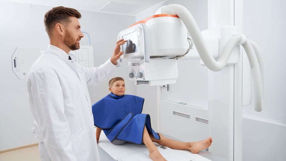
This is particularly important when considering that imaging departments can be intimidating for both children and their caretakers, with large imaging equipment, multiple healthcare professionals, and a busy, noisy environment. As also observed in some adults, children may experience fear or anxiety even more significantly, which can compromise imaging success. This applies especially to young children, who might not fully understand the procedure or the rationale behind it.
While patient trust and cooperation are always important in medicine, they are especially critical in paediatrics. It’s important to help young patients and their loved ones navigate the radiological examination, empowering them to make informed decisions for their health. Equally, it’s crucial to earn the child’s confidence and cooperation both before and during the examination, ensuring a calmer, more supportive environment, which improves the accuracy of results and overall experience.
Supporting the next generation through advanced imaging
By prioritising gentler, patient-focused approaches, the field of paediatric MRI has seen significant advances, while its use in children has expanded rapidly over the past decade, with greater uptake across a broadening range of clinical indications.[15]
A key success factor to further drive more patient-focused innovation for young patients is the integration of paediatric considerations earlier in the clinical development process. There are substantial regulatory efforts to accelerate development and availability of paediatric medication.[16] However, simultaneous submissions for adult and paediatric indications are still uncommon, sometimes resulting in a significant lag before children can benefit from new treatments and technologies. This means paediatric teams often rely on off-label use of newly approved drugs to treat newborns, children, and adolescents before a safety and efficacy assessment of the same rigour as the adult indication for the drug has been made. Therefore, it is important to push for smarter, earlier data-generation strategies in paediatric imaging, from birth to adulthood and across a diverse range of patient populations. These insights are key to bridge the efficacy determined in clinical trials in adults.
Looking ahead, I believe the true promise of continued innovation in paediatric imaging is including children at the earliest possible stage of clinical development. By combining early, data-driven approaches and child-centred care, clinicians and researchers can anticipate paediatric needs, design evidence-based dosing, and support more effective use of imaging agents and therapies. This, alongside a caring and trust-based approach to radiology in paediatrics, will help to transform patient care from an early age.
References
[1] Hussain S, Mubeen I, Ullah N, et al. Modern diagnostic imaging technique applications and risk factors in the medical field: a review. Biomed Res Int. 2022;5164970. https://pmc.ncbi.nlm.nih.gov/articles/PMC9192206/. Accessed December 2025.
[2] O’Connor M, Grassick E. A retrospective analysis of paediatric medical imaging utilisation in Ireland, 2019-2023. J Med Imaging Radiat Sci. 2025;56:102083. https://www.sciencedirect.com/science/article/pii/S1939865425002322. Accessed December 2025.
[3] Thukral BB. Problems and preferences in pediatric imaging. Indian J Radiol Imaging. 2015;25(4):359-364. https://pmc.ncbi.nlm.nih.gov/articles/PMC4693383/. Accessed December 2025.
[4] Simcock, Clare M et al. “Tips for success, managing the paediatric patient in radiology.” Pediatric radiology vol. 55,3 (2025): 411-419. doi:10.1007/s00247-024-06029-w. Available at: https://pubmed.ncbi.nlm.nih.gov/39196382/. Accessed December 2025.
[5] Portelli, Jonathan L et al. “Frequency of paediatric medical imaging examinations performed at a European teaching hospital over a 7-year period.” European radiology vol. 26,12 (2016): 4221-4230. doi:10.1007/s00330-016-4305-7. Available at: https://pubmed.ncbi.nlm.nih.gov/27011372/ . Accessed December 2025.
[6] German Patent and Trade Mark Office. 130 Years Ago: Discovery of X-Rays. https://www.dpma.de/english/our_office/publications/milestones/inventionsthatmadehistory/roentgensx-rays/index.html. Accessed December 2025.
[7] European Society of Radiology. The Gentle Way. The Art of Paediatric Imaging. chrome-extension://efaidnbmnnnibpcajpcglclefindmkaj/https://www.myesr.link/IDoR/IDoR2015_Paediatric%20Imaging%20Book_FINAL.pdf. Accessed December 2025.
[8] Pear, B L. “1896: the first year of X-rays in Colorado.” AJR. American journal of roentgenology vol. 165,5 (1995): 1075-8. doi:10.2214/ajr.165.5.7572480. https://pubmed.ncbi.nlm.nih.gov/7572480/. Accessed December 2025.
[9] Applegate, K. “Image Gently: A campaign to promote radiation protection for children worldwide”. South African Journal of Radiology. 19, 2. https://doi.org/10.4102/sajr.v19i2.919. Available at: https://sajr.org.za/index.php/sajr/article/view/919. Accessed December 2025.
[10] Pollatou, Angeliki et al. “An ode to fetal, infant, and toddler neuroimaging: Chronicling early clinical to research applications with MRI, and an introduction to an academic society connecting the field.” Developmental cognitive neuroscience vol. 54 (2022): 101083. doi:10.1016/j.dcn.2022.101083, Available at: https://pmc.ncbi.nlm.nih.gov/articles/PMC8861425/. Accessed December 2025.
[11] Omics Online. Radiology in Pediatric Imaging Advancements, Challenges, and Clinical Applications. Available at: https://www.omicsonline.org/open-access/radiology-in-pediatric-imaging-advancements-challenges-and-clinical-applications-134712.html. Accessed December 2025.
[12] Lohrke, Jessica et al. “25 Years of Contrast-Enhanced MRI: Developments, Current Challenges and Future Perspectives.” Advances in therapy vol. 33,1 (2016): 1-28. doi:10.1007/s12325-015-0275-4.
[13] Bhargava R, Hahn G, Hirsch AW, et al. Contrast-enhanced magnetic resonance imaging in pediatric patients: review and recommendations for current practice. Magn Reson Insights. 2013;6:95-111. doi: 10.4137/MRI.S12561. https://pmc.ncbi.nlm.nih.gov/articles/PMC4089734/ . Accessed December 2025.
[14] Gajjar, Amar et al. “Pediatric Central Nervous System Cancers, Version 2.2025, NCCN Clinical Practice Guidelines In Oncology.” Journal of the National Comprehensive Cancer Network : JNCCN vol. 23,3 (2025): 113-130. doi:10.6004/jnccn.2025.0012. Available at: https://pubmed.ncbi.nlm.nih.gov/40073837/. Accessed December 2025.
[15] Gallo-Bernal, Sebastian et al. “Whole-Body MRI in Children: Concepts and Controversies-AJR Expert Panel Narrative Review.” AJR. American journal of roentgenology vol. 225,1 (2025): e2432178. doi:10.2214/AJR.24.32178. Available at: https://pubmed.ncbi.nlm.nih.gov/39772586/. Accessed December 2025.
[16] Tanaudommongkon, Irin et al. “Combined Pediatric and Adult Trials Submitted to the US Food and Drug Administration 2012-2018.” Clinical pharmacology and therapeutics vol. 108,5 (2020): 1018-1025. doi:10.1002/cpt.1886. Available at: https://pmc.ncbi.nlm.nih.gov/articles/PMC7572516/?utm_source. Accessed December 2025.
About the author
 Dr Konstanze Diefenbach has held the role of head of radiology research & development at Bayer since 2022. She is a medical doctor and has been trained at the Charité University Hospital Berlin, where she was board-certified in clinical pharmacology. Dr Diefenbach started her career in the pharmaceutical industry as management trainee at the interface of research and development in Schering AG in 2005. Since then, she has gained profound expertise and experience in R&D, as well as medical affairs in various roles, therapeutic areas, and regions including Germany, US, China, and Japan.