Can in vivo imaging deliver the full potential of immunotherapy?
In the quest to control cancer we now face the challenge of translating our increasing insights into the cancer-immunity cycle into more effective immune therapies. In vivo imaging in preclinical models has been instrumental in understanding the complex interactions of cancer and the immune system. The great task now lies in imaging immune responses in real cancer patients in a safe and non-invasive manner, to realise the full potential of immunotherapy in the clinic.
Immunotherapy has shown promising outcomes in multiple cancer types. The Food and Drug Administration (FDA) and European Medicines Agency (EMA) have approved several immunotherapeutic drugs including the checkpoint inhibitors which have been fully accepted in clinical practice. The best-known target for immune checkpoint inhibition is the programmed cell death receptor 1 (PD-1/CD279). It is targeted by the monoclonal antibody drugs pembrolizumab and nivolumab for squamous cell lung cancer, Hodgkin’s lymphoma, cervical cancer, recurrent head and neck squamous cell carcinoma and an ever-broadening range of cancer types.
Atezolizumab, durvalumab and avelumab target the PD-1 ligand (PD-L1/CD274). Atezolizumab is approved for treatment of triple negative breast cancer, both non-small cell and small cell lung cancers and unresectable or metastatic urothelial carcinoma. Durvalumab is approved for metastatic non-small cell lung carcinoma and metastatic urothelial carcinoma. Avelumab is also approved for Merkel cell carcinoma, urothelial carcinoma and renal cell carcinoma (in combination with axitinib). The first checkpoint inhibitor developed was ipilimumab, which targets the cytotoxic T lymphocyte associated antigen 4 (CTLA-4/CD152) receptor. It is approved for metastatic melanoma and more recently also for advanced renal cell carcinoma and mismatch repair deficient metastatic colorectal cancer, but it is less widely used due to a less favourable side effect profile. Both the PD-1/PD-L1 inhibitors can be used in combination with the CTLA-inhibitor, which enhances efficacy but also adds to the side effects.
The question remains when is it beneficial to add ipilimumab to the treatment? Imaging of immune responses to treatments and visualising location and activity of T cells and other immune effector cells, ideally in human cancer patients, can help to answer this question.
Checkpoint inhibitors are not the only immunotherapies that have moved into clinical use and created major advances in cancer treatment. What is clear from on-going clinical trials is that numerous new therapies in the immune-oncology space are emerging and will likely be approved in the near future. For example, cell-based therapies, such as chimeric antigen receptor (CAR) T cells against the CD19 antigen have reached the clinic as second line treatment for blood cancers such as B-cell lymphoma (axicabtagene ciloleucel, tisagenlecleucel) and B-cell acute lymphoblastic leukaemia (tisagenlecleucel).
Despite the beneficial effects of immune checkpoint inhibitors and the emergence of cell-based therapies in clinical studies, their low response rates are, as yet, insufficient to unleash the full potential of these therapies. Only a minority of patients within a cancer type respond, although when they do the benefits are meaningful. Activity in clinical studies seeking to find more effective (combination) treatments has intensified greatly as a result.
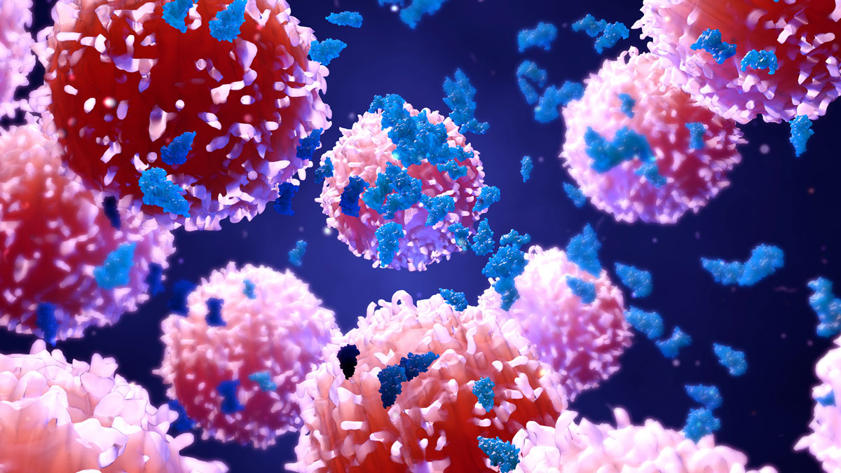
The main rationale for immunotherapies is to induce or enhance infiltration of cytotoxic T lymphocytes (CTL) into the tumour. As one key opinion leader, Professor Tony Ribas, has stated “The T-cell is the drug”. Therefore, in vivo imaging technology could interrogate immunotherapy-induced T-cell responses in cancer patients. It represents a powerful tool to boost further development of immunotherapy without it being too invasive, a drawback of biopsies.
In recent years, the cancer-immunity cycle has been introduced as a concept to describe the necessary steps for effective anti-cancer immune responses. This cycle initiates at the inflamed tumour sites where activated antigen presenting cells (APC) in the tissue recognise and envelope antigenic tumour fragments before migrating to secondary lymphoid organs (SLO).
Upon antigen-specific recognition and activation, T-cells clonally expand and leave the SLO and home in to the tumour sites. T-cells start to infiltrate the tumour microenvironment where antigen-specific T-cells can execute their cytotoxic effector functions. In effective anti-cancer immune responses the subsequent inflammatory response results in the induction of a new wave of effector cells, thereby continuing the cancer immunity cycle.
A variety of different imaging approaches, spanning molecular to whole-body scans, have contributed greatly to our understanding of the conditions required for an effective anti-cancer immune response. Imaging modalities as diverse as intravital fluorescence microscopy and planar bioluminescence imaging, yielded vast amounts of valuable data as molecules and cells were studied in spatio-temporal manners and complemented by more population-based technologies such as flow cytometry. The real breakthrough is the ability to use whole-body medical imaging modalities with relevance for clinical studies which provide tools to study T-cell responses in vivo in a non-invasive manner, showing their induction in the SLO as well as infiltration of tumours for real time activity measures in the tumour microenvironment.
Current clinical research tools, e.g. tumour biopsies and blood-based assays, provide only snapshots of the interactions in the complex human tumour microenvironment following immunotherapy. In many patients, serial and multiple biopsy to determine T-cell engagement is not an option, and blood assay only indicates activation of the immune response.
Imaging tools should focus on accurately reflecting the presence of specific immune cell populations such a CD8 T-cells, which are targeted by the novel immunotherapeutics or combination treatments in a dynamic fashion. Whole body, in vivo imaging has the ability to be used in many scenarios, e.g.:
- Prediction of response or non-response to immunotherapy
- Identify on-treatment responders vs. non-responders to immunotherapy
- Assessment of response to immunotherapy in ambiguous or suspected progressive disease
These types of assessments give us a tool for more efficient immunotherapy development, in vivo imaging should increase our understanding of the critical factors underlying success or failure of immunotherapy in cancer and hopefully drive another step change in development of more effective immune therapies for cancer, helping researchers find new immunotherapy drug classes, optimal treatment combinations and treatment regimens.
About the author
Ian Wilson is chief executive officer of ImaginAb. He was formerly CEO and CTO of Edinburgh Molecular Imaging, leading the development of novel optical imaging agents to improve cancer detection and surgery, and lung disease. He has also held several roles at GE Healthcare between 1996 and 2013, including portfolio and strategy manager and head of biology.