Improving treatment for high-risk cataract surgery
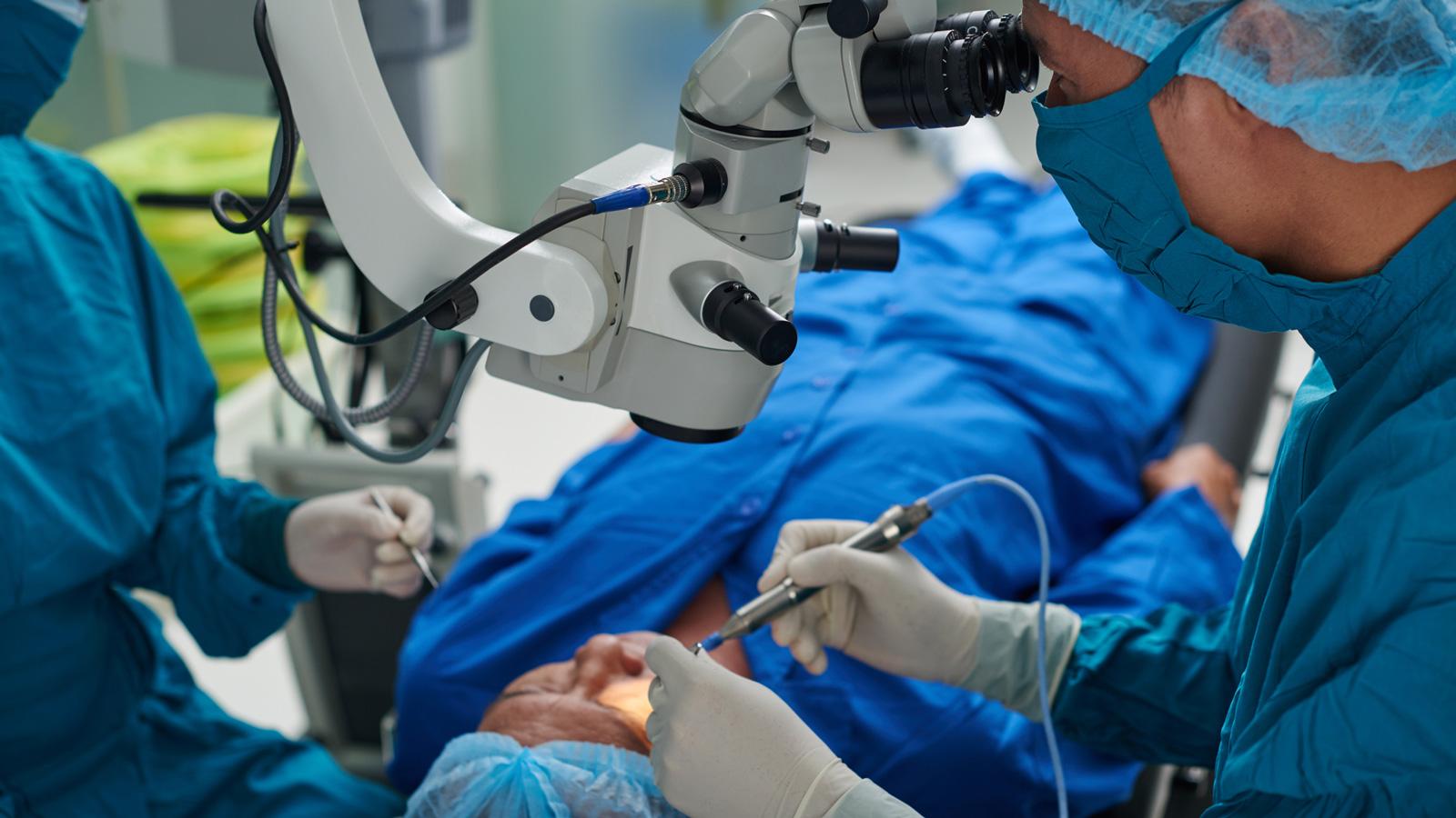
Cataracts rank among the most prevalent age-related ailments. The condition, which is caused by the degradation of proteins in the lens of our eyes, creates a progressively opaque mass in the centre of a person’s vision, blurring, and obscuring vision. While cataracts can cause blindness if left untreated, they can usually be removed using a straightforward and widely-available surgical procedure. In the United States alone, over 4.5 million people undergo this operation annually with remarkable success.1
For a small subset of these patients, however, surgery introduces a new challenge. In people with mature, highly-developed cataracts, the lens becomes extremely hard and inflexible, and breaking it up for removal can cause collateral damage to sensitive endothelial tissue that lines the back side of the cornea. This tissue is normally slow to heal, meaning that added insult from surgery will increase recovery times even in healthy individuals.
For patients that already have chronic eye problems, like diabetic retinopathy or Fuchs’ Dystrophy, a condition in which endothelial cells prematurely die off, negative outcomes from surgery can be even more common. Once damaged, their corneal tissue can take even longer to repair itself, leading to excess swelling and the formation of fluid oedemas in corneal tissue that bend and blur incoming light. Once these oedemas form, vision is dramatically impaired in the patient. Fixing the problem often requires long-term follow-up treatments; the treatments and impaired vision hinder daily activities. In a worst-case scenario, it can require a complete corneal transplant, forcing patients to take immunosuppressant drugs for the rest of their lives.
A problem of cell death
Part of the reason that corneal cells are slow to heal after cataract surgery is due in part to a process called ferroptosis, a newly-identified process of cell death that is associated with oxidative stress. When sensitive eye tissues are disturbed during the operation, reactive oxygen species (ROS) accumulate nearby, bind with the lipid membranes of corneal cells, and, eventually, react with iron inside the cell. This process hinders the cells’ ability to neutralise oxidative molecules, while allowing excess iron to accumulate inside of them. The resulting biochemical reactions can damage or kill the cell.
Usually, the eye initiates a healing response when cells come into contact with Fibroblast Growth Factor 1 (FGF1), a key catalyst for cell division in the cornea. FGF1's primary role is to safeguard cells against damage, including oxidative stress, and promote tissue healing. Unfortunately, native FGF1 is highly unstable and has a short duration of biological activity, meaning that it is not always able to stop the process of ferroptosis once it begins.
Theoretically, increasing the concentration of FGF1 in the eye would enhance healing of corneal oedema and prevent ferroptosis. Several research groups have explored therapeutic versions of the protein for use in treating dermal wounds, yet these efforts have been hindered by the molecule's instability and short half-life. Due to its propensity for breakdown, using FGF1 to stimulate corneal growth would necessitate near-daily injections into the eye, a procedure that few patients would be willing to endure.
Kickstarting the healing process
Trefoil Therapeutics was founded to explore the clinical potential for treating at-risk patients. Despite the challenges of delivering FGF1 into the eye, we believe this growth factor’s ability to kickstart healing in corneal endothelial tissue is a tantalising starting point. By artificially altering the molecule so it becomes more robust and stable, it may be possible to keep FGF1 in the eye for longer periods of time, making it more available to receptors on corneal endothelial cells.
The potential for this sort of protein enhancement is not theoretical. Researchers at Trefoil have already mapped the three-dimensional structure of FGF1, revealing weak points in its molecule. Certain regions of the protein that contain an amino acid called cysteine, for instance, are prone to oxidation, which causes the carefully folded structure of the protein to unravel and fragment. Using existing RNA editing technologies, we believe it is feasible to construct an FGF1-like protein without these oxidation points, making it more persistent in the eye and more readily able to trigger cell division in endothelial tissue.
Enhanced growth factors like this one would give a naturally-occurring molecule a major boost in efficacy, despite sub-optimal conditions in the eye. This could have a major impact on healing in all corneal tissues: when used externally, a modified growth factor has the potential to alleviate excess pain and reduce the risk of long-term harm by speeding recovery from abrasion and other external corneal damage. Likewise, intracameral injection of a modified FGF1 molecule would allow such a drug to target endothelial cells directly, speeding their division and overcoming many of the roadblocks to healing caused by existing pathology.
We firmly believe that the future of post-surgical cataract care will be in harnessing and strengthening the power of natural healing factors. If successful, this sort of medical advancement would give patients with few options for treatment a new chance to regain healthy vision.
About the author
David Eveleth holds the position of president and CEO at Trefoil Therapeutics, a clinical-stage biotech company dedicated to restoring vision in individuals affected by corneal disease.