AI system beats radiologists at predicting lung cancer risk
A deep learning AI system was able to detect malignant lung nodules better than radiologists in a new study by Google and Northwestern University.
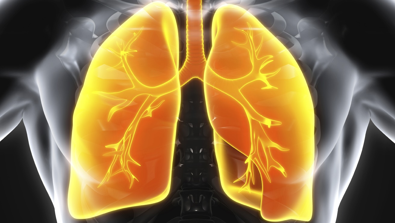
The system analyses low-dose chest computed tomography (LDCT) scans. It provides an automated image evaluation system to enhance the accuracy of early lung cancer diagnosis that could lead to earlier treatment.
The programme utilises both the primary CT scan and, whenever available, a prior CT scan from the patient as input. Prior CT scans are useful in predicting lung cancer malignancy risk because the growth rate of suspicious lung nodules can be indicative of malignancy.
The computer was trained using fully de-identified, biopsy-confirmed low-dose chest CT scans.
It identifies both a region of interest and whether the region has a high likelihood of lung cancer.
The model outperformed six radiologists when previous CT imaging was not available and performed as well as the radiologists when there was prior imaging.
The system also produced fewer false positives and fewer false negatives, which could lead to fewer unnecessary follow-up procedures and fewer missed tumours if it were used in a clinical setting.
Google scientists developed the deep-learning model and applied it to 6,716 de-identified CT scan sets provided by Northwestern Medicine to validate the accuracy of its new system.
The scientists found the artificial-intelligence-powered system was able to spot sometimes-minuscule malignant lung nodules with a model area under the curve (AUC) of 0.94 test cases.
“Radiologists generally examine hundreds of two-dimensional images or 'slices' in a single CT scan but this new machine learning system views the lungs in a huge, single three-dimensional image,” said study co-author Dr Mozziyar Etemadi, a research assistant professor of anesthesiology at Northwestern University Feinberg School of Medicine and of engineering at McCormick School of Engineering.
“AI in 3D can be much more sensitive in its ability to detect early lung cancer than the human eye looking at 2D images. This is technically '4D' because it is not only looking at one CT scan, but two (the current and prior scan) over time.”
Etemadi added: “The system can categorise a lesion with more specificity. Not only can we better diagnose someone with cancer, we can also say if someone doesn't have cancer, potentially saving them from an invasive, costly and risky lung biopsy.”
The paper was published in Nature Medicine.